Summary:
In order to segment the lung parenchymal region from the chest region containing background and noise, first, the traditional regional growth method is used to preliminarily locate the boundary contour of the lung; secondly, the lung boundary noise is removed and the lung boundary is repaired by the adaptive curvature threshold method. Finally, the DRLSE model in the level set method is used to accurately segment the lung region. Integrate the two methods to segment the lung area, which effectively prevents the image edge from being missed and can process lung images of various types of lesions. Among the 150 randomly selected images, the accuracy of segmentation is 96.9%, and the time taken to segment an image is about 0.72 s, which has strong robustness and high segmentation accuracy. The algorithm can accurately and completely segment the lung area and retain details in the lung area.
0 Preface
With the deteriorating atmospheric environment, the incidence of lung diseases has increased year by year. According to statistics, lung cancer accounts for up to 21% of cancer patients and the mortality rate remains high. Clinically, the cure rate of early-stage lung cancer is as high as 90%. Early detection of abnormal lung conditions can control the condition and reduce mortality. At present, mainly by observing the lung CT sequence images to find the lesion information, accurately segmenting the lung region is the key precondition for locating the tumor. For the segmentation of lung regions, experts and scholars at home and abroad mainly implement the threshold method, regional method, genetic method, level set method and artificial neural network.
Threshold method is the most commonly used and simple method of segmentation. The principle is to draw the gray histogram of the image. By selecting the threshold in the histogram, the image is classified and the segmentation result is obtained. In [1], Guo Shengwen et al. proposed to first apply the adaptive threshold method to binarize the lung region image, and then use the feature classifier to accurately segment the lung region. The threshold method requires that the gray value of the image be uniformly distributed, and the peaks and valleys can be clearly observed in the histogram, otherwise the specified region cannot be accurately segmented, and the limitation is relatively large. In [2], Gao Guorong et al. proposed the regional growth method to segment the lung region. According to the set seed point, the high density information such as trachea and blood vessels in the adhesion lung was accurately removed, but the seed points need to be manually selected. Large, practical applications need to combine other algorithms to segment the results better. In [3], Qin Xiaohong et al. proposed the use of a global optimization capability strategy in genetic algorithms to achieve segmentation of the lung region. This algorithm can simplify segmentation and improve the search scope, but it needs to provide a large number of training samples and the extraction of features also requires It takes a lot of time, convergence speed is slow, and practicality is not high. In [4]-[5], Wei Ying et al. used the CV level set method in the Geometric Active Contour (CAC) method to segment the lung region, and combined the lung structural features to improve the CV method. The accuracy and accuracy of partial boundary segmentation have improved.
Compared with the traditional GAC model, the segmentation accuracy of the method is higher, the convergence speed is faster, and the leakage of the edge is also effectively prevented. In summary, the level set method developed by the active contour model has better segmentation performance and lower algorithm complexity, and it can also handle complex images. Therefore, this paper combines traditional segmentation methods with modern segmentation methods and proposes a segmentation of pulmonary CT images based on region growing method and level set. First, an adaptive thresholding method is used to binarize CT images of the lungs, and then a regional growth method is used to perform rough segmentation on the lung regions [6]. Finally, the geometric active contour model in the level set method is used to achieve accurate segmentation. After the simulation test on the MATLAB software platform, the experimental results show that the segmentation effect of the CT image segmentation in the lungs is better by using this method.
1 algorithm principle
1.1 Threshold Method Principle
The threshold method [7] is a region segmentation technique that divides a gray value into two or more gray ranges, selects one or more appropriate thresholds, and judges whether or not an area satisfies a threshold according to the difference between the target and the background. Separate the background and the target to produce a binary image. There are two forms of thresholding: global thresholds and adaptive thresholds. Only one threshold is set for the global threshold. The adaptive threshold sets multiple thresholds, and the target and background regions are segmented by determining thresholds at the peaks and valleys of the grayscale histogram.
Whether the threshold selection is appropriate or not is determined and the image segmentation is determined to be good or bad. In this paper, an adaptive threshold method is used to binarize the lung image. The structure of the lung region is complex. Under different environments and conditions, the image performance is also different. Applying an adaptive threshold method and dynamically selecting multiple thresholds can achieve better binarization of the lung region.
This article uses the popular OSTU algorithm to select the threshold. The basic principle is: using the threshold to divide the histogram into two parts, and when the two parts are divided into the largest variance, the optimal threshold is obtained [8]. The adaptive threshold method finally transforms the DICOM-format medical CT image into a black-and-white binary image so that the image can be segmented, extracted, and recognized later.
1.2 principle of regional growth method
The basic idea of ​​the region growing method is to synthesize a pixel or subregion into a larger region according to a predefined growth criterion. The basic processing method is to form a growth region starting from a set of “seed†points, ie, those predefined attributes ( For example, pixels of a gray level or color similar to seed points are attached to each seed point [9]. The key issue of regional growth is the selection of similarity criteria and the setting of stopping rules in regional growth. The criterion can be set by the similarity of the brightness value, texture, and color of the image. When no more pixels satisfy the criteria contained in the area, the process of growing the area stops. The segmentation method based on region growing is simple in calculation and can realize the initial segmentation of the lung region and obtain the initial contour information of the region.
1.3 Level set principle
The level set method is the Geomet Active Contour (GAC) model in Active Contour (AC), which is an image segmentation method developed in recent years. The basic idea of ​​level set method for image segmentation is [10]: Through the constant evolution of the curve motion, the boundary of the image is always searched until the target contour is found and the movement curve is stopped. The curve needs to move along each three-dimensional slice plane of the CT image. Slices are taken from different levels of the three-dimensional surface to obtain a closed curve for each layer. As time goes by, the horizontal level is changed, and a corresponding shape extraction profile is finally obtained. .
There are three commonly used level-level models, the MS (Mumford-shah) model, the CV (Chan-Vese) model, and the DRLSE (Distance Regularized Level Set Evolution) model [11]. The first two models are the traditional level set models. Is the use of regional statistics to segment, to overcome the use of gradient (edge) information leads to weak edge missed detection, multiple noise sensitive and other issues. However, finding the approximate region requires constant initialization of the level set function, which still has the problems of low computational efficiency and difficulty in implementation. The DRLSE model has improved the traditional method by adding a constraint to the level set function, avoiding the periodic initialization of the level set function, improving the convergence speed, and reducing the running time. According to the analysis of the above model, the DRLSE model was used to extract the lung contour region.
2 Segmentation of lung CT images with regional growth and level integration
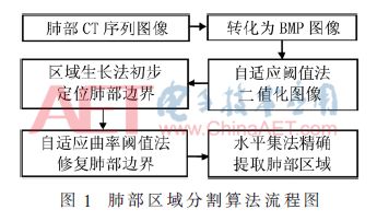
After obtaining the DICOM format CT images, the work of this paper is to accurately segment the complete lung parenchymal area to facilitate the subsequent diagnosis and quantitative data analysis of lung diseases. The segmentation of the lung region is divided into four parts: image binarization, regional growth method to initially locate the lung border region, and lung boundary repair, and the level set method accurately extracts the outline of the lung region. Figure 1 shows a flowchart of the algorithm for segmenting lung regions.
2.1 Image Preprocessing
The collected DICOM format image cannot be displayed on the computer screen and must be converted to BMP format. Because the storage modes of the two image formats are different, the vertical mirroring transformation is applied to format conversion in this paper. The medical image vertical mirroring formula is expressed as:

G0(-n0, k0) represents the pixel value of the pixel point (-n0, k0) in the DICOM format original image I0, and Gvm(n0, k0) represents the pixel value of the pixel point in the BMP format image Ivm after the vertical mirroring.
2.2 OSTU Image Binarization
The OSTU method is an automatic threshold segmentation method. According to the threshold, the image region is divided into two groups. When the variance of the two regions divided reaches the maximum, the image segmentation is performed by obtaining the optimal segmentation threshold. It belongs to single-threshold segmentation and divides the image into two types, the background and the target. After processing, the grayscale image is converted into a black and white image. The processing steps are as follows:
(1) Read lung CT images and convert them to BMP format images.
(2) Use the imhist function in the MATLAB software toolbox to draw a grayscale histogram of the image. The formula is:

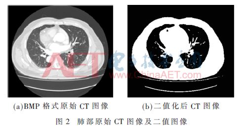
Among them, I is the original image, n is the gray level, and M is the grayscale histogram of the image I. According to the difference in the gray level between the lung region and the surrounding organs, the overlap region in the histogram is minimized, the maximum class difference between the two regions is obtained, the best segmentation threshold is obtained, and the lung region is segmented as a target region according to the threshold value. As the background area in other areas, the image after binarization using the OSTU method is shown in FIG. 2 .
2.3 Regional Approach to Preliminarily Locate Lung Borders
Binarized lung CT images did not accurately separate the lung parenchyma from the background of the image. The edge information of the lung parenchyma was also confounded in the background region. At the same time, the lung parenchyma region contained some noise such as trachea and air. In this paper, the region growing method is used to locate the lung region boundary and eliminate interference information such as noise.
The regional growth method needs to solve three problems: (1) It is necessary to determine a certain pixel in the region as the growth seed point; (2) to determine the growth criterion of the seed point; and (3) to determine the termination condition of the seed point in the growth process.
2.3.1 Preliminary Positioning of the Lung Region Boundary
The steps for locating the lung boundary algorithm are as follows:
(1) Enter the binarized image and calculate the maximum resolution Amax, Bmax in the X and Y axis directions on the image.
(2) Select the target region R, calculate the average gray value Tm of the target region, and set there are N pixels in the region, then the gray average of the region is:
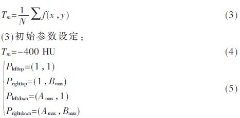
(4) Selection of seed points: Set the four-neighborhood template from the far left of the image of the target area and scan the surrounding pixels. If the gray value of the pixel is less than Tm, it is the seed point.
(5) Set the region growth criterion T: where I(Ai, Bi) is an arbitrary pixel value of the original image, and N4(Ai, Bi) is a four-neighborhood pixel point of Ai, Bi. The four-neighborhood pixel points are compared with the grayscale average.

The target area is scanned from the upper left to the lower right, and all the pixels satisfying equations (6) to (8) are found.
(6) Search for the four-neighborhood pixels around all the seed points that meet the criteria until the condition is not satisfied and the segmentation is completed.
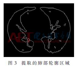
The regional growth method can be used to initially locate the contour region of the lung parenchyma, and the result of extracting the contour region is shown in FIG. 3 .
2.3.2 Trachea removal
The algorithm idea for extracting the external trachea of ​​the lung parenchyma is: after setting the seed point, a checkerboard distance marking method is used to generate the pixels in the region, and the distance K between the checkerboards is determined. After the Nth iteration, according to the region growing rule, the search satisfies the conditions. All the pixels were finally set to the growth termination condition and the trachea area was detected. Let V(K) be the area value that satisfies the growth of the region, plot the V(K) curve, and observe the change in the median value of the graph. If the value suddenly increases, it indicates that the tubular organ is detected and the region is extracted and removed. Specific steps are as follows:
(1) Selection of seed points: Search for seed points from the middle lung field slice, set a 4×4 template, search for all the areas that satisfy the pixel points, and use the area growth method to obtain seed points.
(2) Growth rule: Search for the value of the four neighboring pixels around the seed point, keep the pixel value between T+4 and T-4, and search for an area of ​​not less than 100 pixels.
(3) Termination condition: Calculate the area derivative of the function V(K). If the area derivative value is greater than zero, stop the search, extract the trachea region, and obtain the lung parenchymal region without interference information.
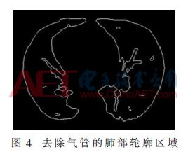
The lung region of the trachea was removed as shown in FIG.
2.4 Pulmonary edge repair
A large number of trachea, blood vessels, etc. are distributed on the border of the lungs. At the same time when the trachea is removed, the border area is uneven, and the edges need to be repaired to obtain a smooth and complete outline of lung parenchyma.
2.4.1 Curve smoothing
Before the restoration of the lung boundary, the boundary must be smoothed. Otherwise, the parameters cannot be accurately and automatically calculated to calculate the curvature of the curve, and the boundary can be accurately repaired.
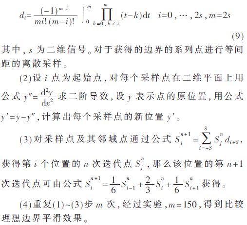
This article applies iterative sampling point algorithm to smooth the lung contour curve. Extract the contour area of ​​the lungs, extract a series of points {i} on the edge, and obtain the concave and convex areas of the series point connecting lines. Apply the Newton-Cuttings formula to the concave and convex points for the concave and convex areas. After many iterations, make the series of edge points. The process of approximating a linear relationship to achieve smooth edges. The algorithm implementation steps are:
(1) Set the step length λ = 0.9, according to the Newton-Cutter formula:
2.4.2 Boundary Repair
In this paper, the adaptive curvature threshold method is used to repair the lung boundary. After the smooth edge, the curvature threshold of the point can be accurately calculated. The pit with the larger curvature value is the point near the edge of the lung to be repaired. The algorithm steps are as follows:
(1) The curvature threshold of the set point is μ, the boundary area is denoted by P, and the average of the height of the calculated area point  , Let μ=
  , the discrete points of the sampling area P.
(2) Calculate the height of each discrete point i in the area P, set it as ωi, and find all the pit areas. If ωi>μ, delete the point i and connect the neighborhood point of point i.
(3) Repeat steps (1) and (2) until all points in the boundary area have been searched, and the length of the area is the same as the original length.
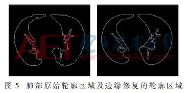
Finally, a smooth and complete lung contour area was obtained. The effect of contrast before and after repair is shown in Fig. 5.
2.5 Level Set Accurate Extraction of Lung Area
This article selected the DRLSE model to accurately extract lung regions. The advantage of this model is that when iterative methods are used to find matching points in the image, it is not necessary to repeatedly initialize the level set function, which improves the speed of operation and reduces the amount of data calculation. The mathematical formula of the level set function φ is expressed as:
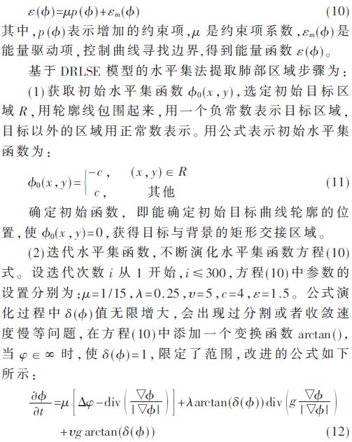
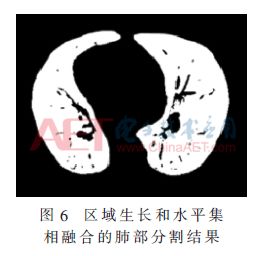
(3) Repeat step (2), and continue to iterate the level set function until satisfactory results are achieved. The value of i does not exceed 300. Stop the iteration to obtain the precise contour area of ​​the lung boundary after the level set iteration, and the boundary curve is also obtained. For good convergence, the extracted lung region image is shown in Figure 6.
3 experimental results
3.1 Sources of data
To verify the feasibility of this method and the accuracy of segmentation, 120 images were randomly selected from the LIDC lung image public database, and 30 images containing lesion information were extracted from a hospital in Baotou. Each image was equipped with expert segmentation. The "gold standard" is also an important criterion for evaluating the quality of the algorithm. After screening and identifying approximately 110 images showing signs of lung lesions, more than 90% of the lung regions were isolated in the lung regions, so no mention was made of left and right lung separation. The image has a resolution of approximately 0.6 mm to 0.8 mm, a size of 512×512, and a scanning layer of approximately 30 layers.
3.2 Experimental Environment
The experiment platform of this experiment is: Windows 7 operating system, i5 processor CPU, 8 GB RAM, programming software is MATLAB R2015a, Visual Studio 2012.
3.3 Results and Analysis
In order to verify the feasibility of image segmentation after the two methods were combined, the segmentation results of two groups of different lung lesion images were listed, as shown in FIG. 7 .
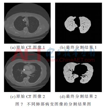
From the segmentation results, we can see that the method can accurately and completely extract the lung parenchyma area, overcome the problem of boundary missed detection and other problems caused by the use of the region growth method alone, and combine the accurate segmentation of the level set method in the later period to clearly observe the lungs. Substantial internal details and more accurate positioning of lung edge information also avoid problems such as over-segmentation of images. At the same time, lung images of various types of lesions can also be processed to lay the foundation for further analysis and processing of subsequent images.
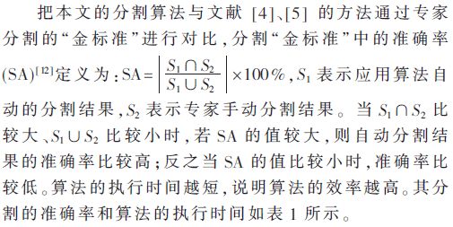
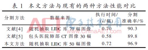
From Table 1, we can see that the algorithm in this paper sacrifices time but improves the segmentation accuracy. From the segmentation results, this algorithm has strong feasibility. The algorithm is automatic computer segmentation, and the time and efficiency are still more time-efficient than semi-automatic or manual segmentation.
4 Conclusion
Segmentation of lung images is a prerequisite for computer-assisted clinicians to detect lung diseases, and the segmentation results directly affect the accuracy of disease detection. This paper presents a lung segmentation method that combines regional growth and level collection. After simulation experiments, the algorithm can effectively and accurately segment the lung area, and retain the internal details of the lung, laying the foundation for the detection and extraction of subsequent pulmonary nodules.
The algorithm of this paper is also inadequate. There is no segmentation of the lung area where the left and right lungs are constricted. In the drawing, two pictures of left and right lung adhesions are removed. Subsequent studies will be conducted to segment the left and right lung adhesions. At the same time, this algorithm can also be applied to the segmentation of lung nodules and pleural nodules to test the generality of the method, and to improve and optimize the problems of segmentation.
8 Inch Tablet
Today let`s talk about 8 inch tablet with android os or windows os. There are many 8 inch tablets on sale you can see at this store. 8 inch Android Tablet is absolutely the No. 1 choice if you are searching a student online learning project. 8 inch windows tablet is more welcome when clients are looking tablet for business application. The most welcome parameters level is 2 32Gb with 4GB lite, 4000mAh, android 11 only around 60usd, price will be much competitive if can take more than 1000pcs. 7 Inch Tablet wifi only, android tablet 10 inch, Amazon 8 inch tablet is also alternative here. Except tablet, Education Laptop, Gaming Laptop , 1650 graphics card laptop, Mini PC and All In One PC are the other important series.
Therefore, you just need to share the configuration, application scenarios, quantity, delivery time, and other special requirements, then will try our best to support you.
Any other thing in China we can do, you can also feel free to contact us.
8 Inch Tablet,8 Inch Android Tablet,Amazon 8 Inch Tablet,8 Inch Tablets On Sale,8 Inch Windows Tablet
Henan Shuyi Electronics Co., Ltd. , https://www.shuyilaptop.com